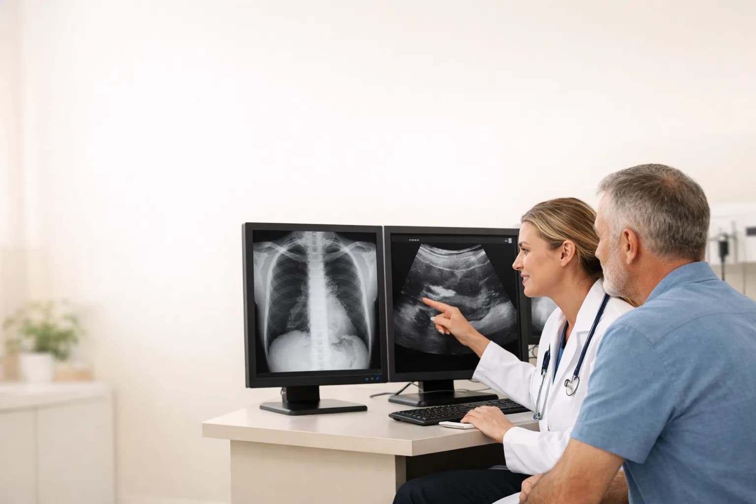
X-rays use ionizing radiation and are strong for many bone and chest questions; ultrasound uses sound waves for real-time soft-tissue imaging. Learn how symptoms, body area, pregnancy, and test limitations guide the choice.
Answer First: X-Ray Favors Dense Structures; Ultrasound Shows Soft Tissue in Motion
X-ray imaging passes a small amount of ionizing radiation through the body. Dense structures such as bone absorb more and appear differently on the image. Ultrasound sends high-frequency sound waves into tissue and builds images from returning echoes, allowing real-time views of many organs, fluids, and blood flow.
NPMD offers both X-ray and diagnostic ultrasound. The provider chooses based on the clinical question rather than which machine seems more advanced.
When X-Ray Is Commonly Useful
X-rays are often the first test for suspected fracture, joint alignment, arthritis, and selected chest symptoms. A chest X-ray can show the lungs, heart silhouette, airways, chest bones, and some fluid or air patterns. It can also document placement of certain medical devices.
A plain X-ray has limits. Small fractures, early infection, ligament injury, cartilage problems, and some tumors may not be visible. A normal image does not always end the evaluation when examination findings or symptoms remain concerning.
When Ultrasound Is Commonly Useful
Ultrasound can evaluate soft-tissue lumps, abdominal and pelvic organs, gallbladder, kidneys, thyroid, pregnancy, some tendons, and blood flow when Doppler is used. It can also guide certain procedures because the image updates in real time.
RadiologyInfo explains that ultrasound uses sound waves, does not use ionizing radiation, and shows soft tissues that may not appear well on X-ray. The exact capability depends on the probe, body area, operator, and clinical question.
Bone, Joint, and Soft-Tissue Injuries
After a fall or impact, X-ray commonly evaluates fracture or dislocation. Ultrasound may help assess selected tendons, muscles, fluid collections, or superficial structures. Neither automatically replaces MRI or CT when a deeper or more complex injury is suspected.
The provider considers where it hurts, mechanism, swelling, weight-bearing, range of motion, nerve symptoms, and examination findings. Severe deformity, open injury, loss of circulation, or major trauma needs emergency care rather than routine outpatient comparison shopping.
Abdominal and Pelvic Symptoms
Ultrasound is often chosen for gallbladder, liver, kidney, pelvic, pregnancy, and vascular questions. Gas and body depth can limit the view. X-ray may be used for selected bowel patterns, foreign bodies, stones, or other questions but cannot fully characterize many soft-tissue organs.
Preparation varies. Some ultrasound exams require fasting; others require a full bladder. Ask for written instructions. Eating, drinking, or emptying the bladder at the wrong time can reduce image quality and may require rescheduling.
Radiation and Pregnancy
Ultrasound does not use ionizing radiation. X-ray does, but medically necessary exams are planned to use the lowest exposure needed for diagnostic images. Tell the ordering clinician and technologist if you are or may be pregnant so benefits, timing, shielding practices, and alternatives can be assessed.
Do not avoid urgent necessary imaging solely from fear, and do not request imaging without a clinical reason. The relevant comparison is the risk of the test versus the risk of missing or delaying the condition being investigated.
What the Appointment Feels Like
X-rays are usually quick. The technologist positions the body and may ask you to hold still or briefly hold your breath. Ultrasound uses gel and a handheld probe pressed over the area; pressure may be uncomfortable when the region is tender.
Remove jewelry or clothing as directed and tell staff about mobility limitations. Bring prior imaging when it was performed elsewhere because comparison can help determine whether a finding is new, stable, improving, or worsening.
Results and Possible Next Steps
A radiologist or qualified clinician interprets the images and creates a report for the ordering provider. The report must be combined with symptoms, examination, and sometimes laboratory results. An incidental finding may be harmless, important, or require another test.
NPMD's diagnostic testing and imaging overview can help you prepare, while sick visits can connect symptoms with an appropriate order. New severe symptoms should not wait for a routine report discussion.
Questions to Ask Before Imaging
What exact question should this test answer?
Ask which body area and diagnosis are being evaluated and what the test may miss.
How should I prepare?
Confirm fasting, bladder, clothing, medication, pregnancy, prior imaging, and when results will be available.
Where can I begin?
Review EKG testing when symptoms are cardiac, then request an evaluation for non-emergency imaging needs.